
Research
Single Atom Electron Spin Resonance
[1] S. Baumann et al., Science 350 (2015)
[2] P. Willke et al., Science Advances 4 (2018)
In 2015, ESR on individual magnetic atoms on surfaces was successfully implemented in a scanning tunneling microscope for the first time[1], manifesting a major step towards a quantum coherent control thereof.
A schematic of the setup is shown in Fig. 1A. A single Fe atom is put on a thin insulator of magnesium oxide (MgO) and imaged by an STM (Fig. 1B). Experiments are conducted at low temperatures (~1 K) and in ultra-high vacuum (UHV). The role of the thin-insulator MgO here is to decouple the atomic spin center from interaction with conduction electrons of the underlying silver substrate which ensures to protect their magnetic properties. The spin states |0> and |1> of the atom are Zeeman-split by a frequency f0 using a macroscopic magnetic field B. In order to perform ESR a radiofrequency (RF) voltage is applied to the STM junction that coherently drives the spin system into resonance. Thus, a change in the state population of the atom can be observed at the resonance frequency f0 (Fig. 1C). We detect the state population here using a spin-sensitive readout provided by a magnetic tip acting as a spin filter [1]. It is prepared by picking up several Fe atoms from the surface, that subsequently form a spin cluster at the tip apex.
Using this technique, ESR measurements were performed on single iron (Fe), copper (Cu) and titanium (Ti) atoms (See below). It is also noteworthy that this technique exceeds the energy resolution of conventional STM spectroscopy (limited by electronic and thermal broadening) by roughly a factor of 1000 reaching ~10 neV energy resolution.
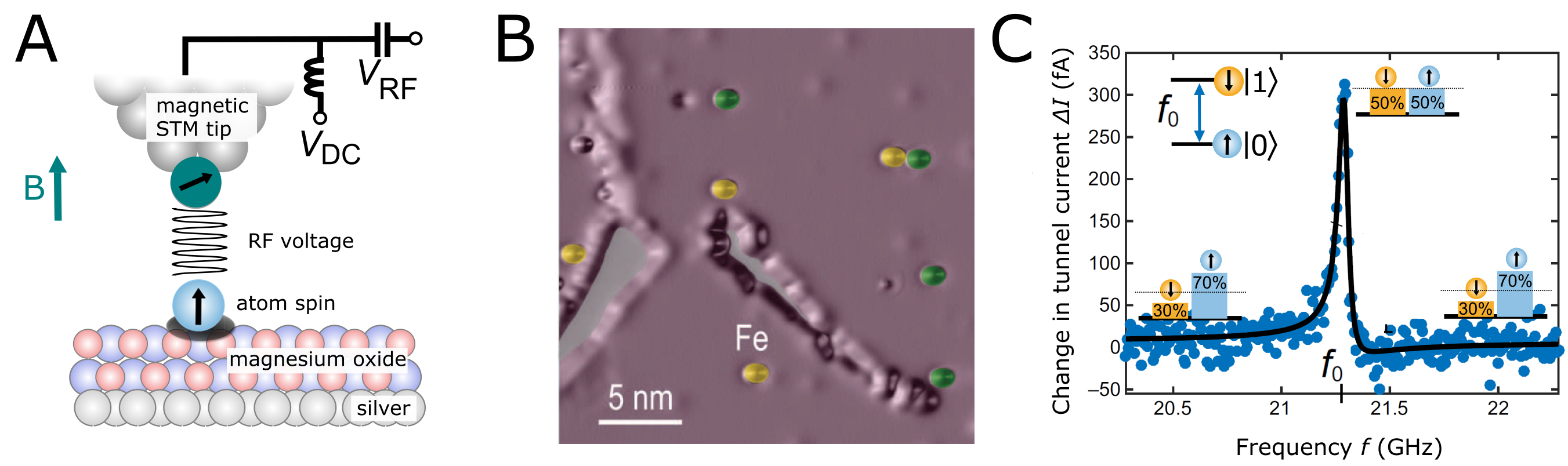
Fig. 1 A, Single atom electron spin resonance. Individual magnetic atoms are deposited on bilayer MgO on top of a silver crystal. A Zeeman split ground state is prepared by a macroscopic B-field. The splitting is typically chosen to yield ~20 GHz. ESR is achieved by introducing a radiofrequency voltage VRF in the tunnel junction in addition to the conventional DC tunnel voltage VDC. B, Constant-current STM image of Fe (yellow) and Co (green) atoms on MgO/Ag[1]. C, ESR spectrum taken on the atom detected by a change in the spin-polarized tunnel current ΔI when the frequency f of the RF voltage is swept. Solid line is a fit to an asymmetric Lorentzian [2]. Insets indicate the change in state population.
Quantum Sensing
[1] T. Choi,…, PW et al., Nat. Nano 12 (2017)
[2] F. Natterer, … PW, et al., Nature 543 (2017)
[3] K. Yang, ... PW, et al., PRL (2017)
[4] P. Willke et al., Nature Physics 15 (2019)
As in conventional ESR, the atom’s resonance frequency f0 is sensitive to any local magnetic field. Therefore, single atoms can be utilized as quantum sensors to probe the magnetic interaction with their environment.
The unprecedented combination of energy and spatial resolution of ESR-STM allowed to study weak magnetic interactions such as magnetic dipolar interaction between atoms on surfaces [1]. A single Fe atom (sensor) can couple to other nearby magnetic atoms (target) when they are separated by only a few nanometers. An ESR spectrum taken on the sensor atom reveals two peaks as shown in Fig. 2. This is caused by thermal fluctuations of the two spin states (up and down) of the target atom. Subsequently, its dipolar magnetic field leads to a frequency splitting Δf of the ESR signal of the sensor atom. The intensity of the two peaks reflects the state population of the ground [P(|0>)] and excited state [P(|1>)] of the target atom. Distance-dependent measurements between target and sensor atom reveal an Δf~r‒3-power law of the peak splitting as predicted by magnetic dipole-dipole interaction [1]. This allows to determine the magnetic moment of the target with high precision (<0.01 μB) and was applied in the following to Fe, Co, Ti and Holmium (Ho) atoms [1], [2], [3]. This technique also allows to estimate the spin relaxation time T1 of the atom. For single Holmium atoms it revealed an extraordinary long spin lifetime on the order of several hours, which realized a long-standing goal for molecular magnets. In addition, it was found that the exchange interaction becomes dominant when the atoms are closer than 1 nm [1][3]. For Ti dimers, this interaction was found to be much more pronounced [1].
In addition, the interaction of atomic spins with the field of the magnetic STM tip can be measured. This effectively shifts the resonance frequency f0 of the atom spin with the position of the STM tip. Therefore, one can take advantage of the STM tip to tune the spin system into resonance establishing magnetic resonance imaging (MRI) on the atomic scale [4]. This allowed us to map the 3D magnetic interaction between a spin center on the surface and the magnetic tip. This approach is very similar to other scanning-field gradient techniques such as magnetic resonance force microscopy or scanning NV-center magnetometry. However, it exceeds the spatial resolution of these methods by 1-2 orders of magnitude and establishes atomic-scale imaging of spin centers with high energy resolution.
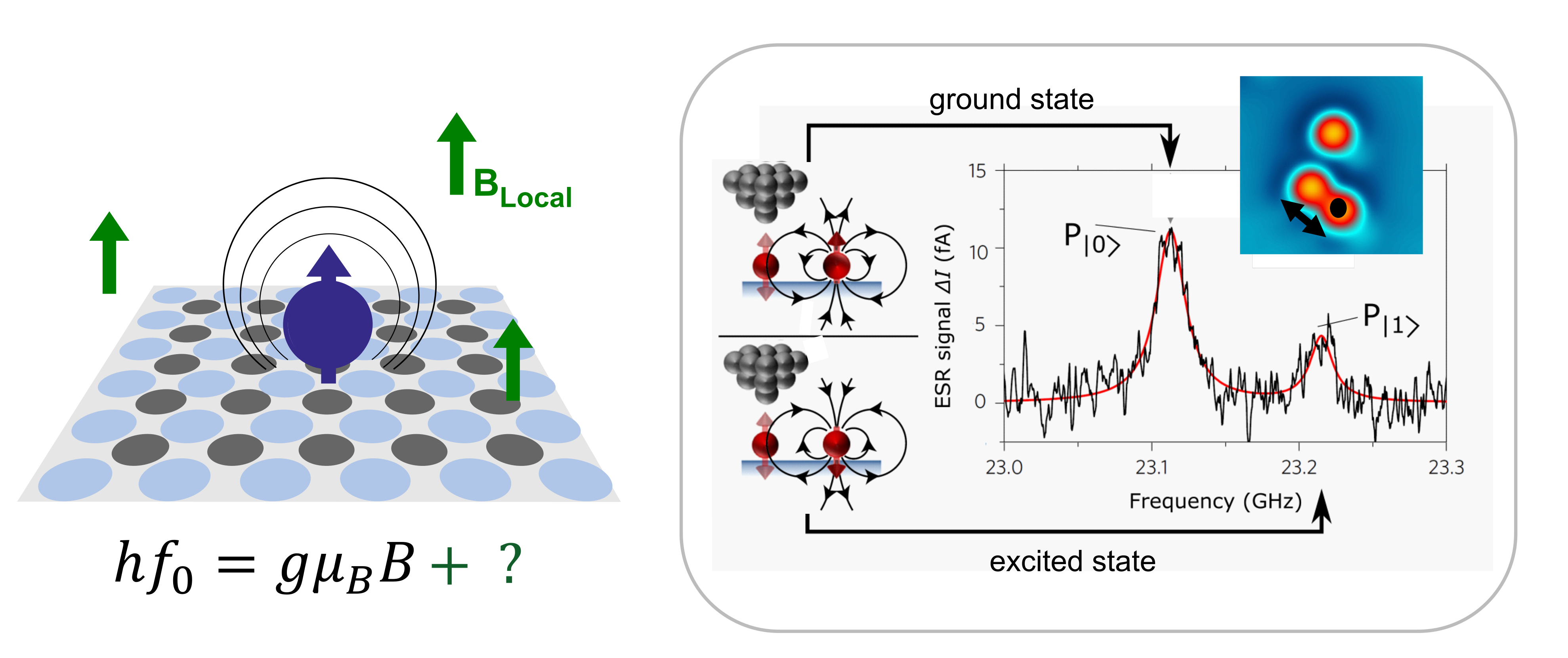
Fig. 2 left: Illustration of a single atom as a quantum sensor. right: ESR spectrum (black curve) of an Fe atom when another atom (target) is positioned ∼2.46 nm away. The two spectral peaks with frequency splitting Δf are caused by magnetic dipole-dipole interaction between the atoms and correspond to the two magnetic states of the target atom, since each state of the target atom generates a different local magnetic field at the sensor atom (sketches to the left) [1].
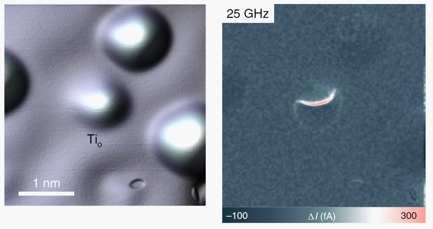
Fig. 3: Magnetic Resonance Imaging of Single Atoms. Left: STM Topography image of a single Titanium atom (center). Right: Change in Tunneling Current in a spatially resolved ESR scan, highlighting the resonant interaction of the atom on the surface and the magnetic STM tip.
Single Atom Nuclear Spins
[1] P. Willke et al., Science 362 (2018)
[2] K. Yang, PW et al. Nature Nano 13 (2018))
Still, the energy resolution of ESR-STM permits to resolve the hyperfine coupling of individual atoms [1][2] (Fe, Ti and Cu, see Fig. 4A and Fig. 4B). This manifests the first detection of single-atom nuclear spins in an STM experiment, allowing to distinguish different isotopes atom by atom. The experiments revealed that the hyperfine spectrum is influenced by the atomic-scale environment of the atom. For instance, atoms could be moved into different binding configurations on the MgO substrate using the STM tip. Subsequently, their ESR spectrum showed a significant change in the hyperfine interaction. This in return permitted to extract atom- and position-dependent information about the electronic structure of the ground state [1]. In addition, we demonstrated that the spin-polarized tunneling current of the STM tip can polarize the nuclear spin by transferring spin angular momentum to the nucleus. This experiment, performed on single Cu atoms, allowed to tune the nuclear spin state by the tunnel current and to initialize it far away from thermal equilibrium (Fig. 4B). The underlying spin transfer torque mechanism relies on spin flip-flop transitions between the tunnel current electrons, the Cu electron spin (S=1/2) and the Cu nuclear spin (I=3/2). Nevertheless, this strong interaction of the nuclear spin with the tunnel current is also responsible for its short spin lifetime. Using rate equation models, we could estimate a nuclear spin lifetime of T1N~500 ns [2].
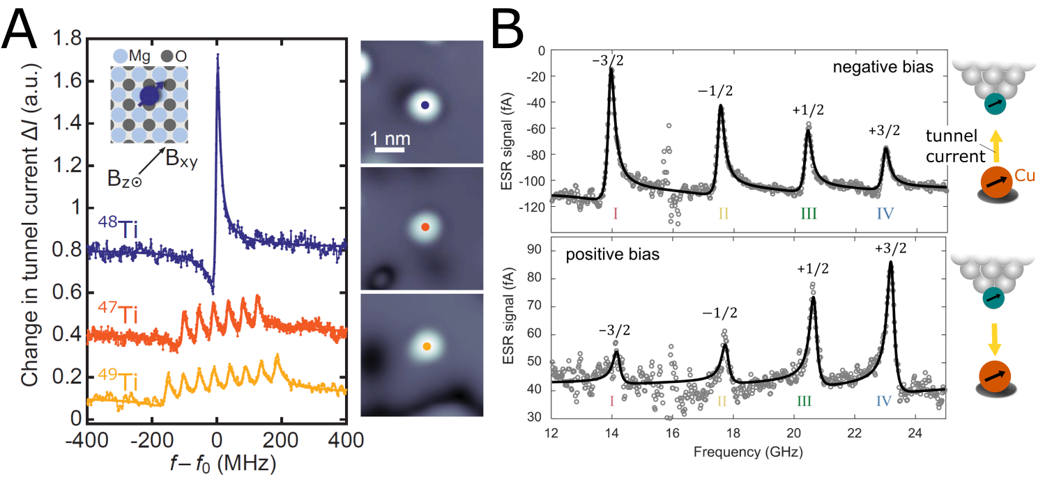
Fig. 4: A, Hyperfine Interaction of Single Atoms. On the left are ESR spectra revealing the hyperfine interaction for individual titanium atoms. Different numbers of peaks are observed for 48Ti (blue, nuclear spin I = 0), 47Ti (orange, I = 5/2) and 49Ti (yellow,I = 7/2) associated with the different possible nuclear spin configurations. On the right are STM images for each atom [1]. B, ESR spectra of a 65Cu atom at VDC= –22 mV and VDC= +22 mV for the same tip reveal a great change in the peak intensity for individual nuclear spin states induced by spin‐transfer torque from the tunnel-current to the nuclear spin (see sketch on the right) [2].
Coherent Spin Manipulation
[1] K. Yang, …, PW et al. Science, 366 (2019)
[2] P. Willke et al., Science Advances 4 (2018)
Using pulsed ESR, coherent manipulation of the single atom spins becomes possible. In Rabi oscillation measurements (Fig. 5A) we observe several coherent oscillations of the electron spin. In Hahn Echo measurements (Fig. 5B), we find a phase coherence time of ~200 ns, mostly limited by nearby tunnel electrons. This was previously found to be the dominating source of noise [2].
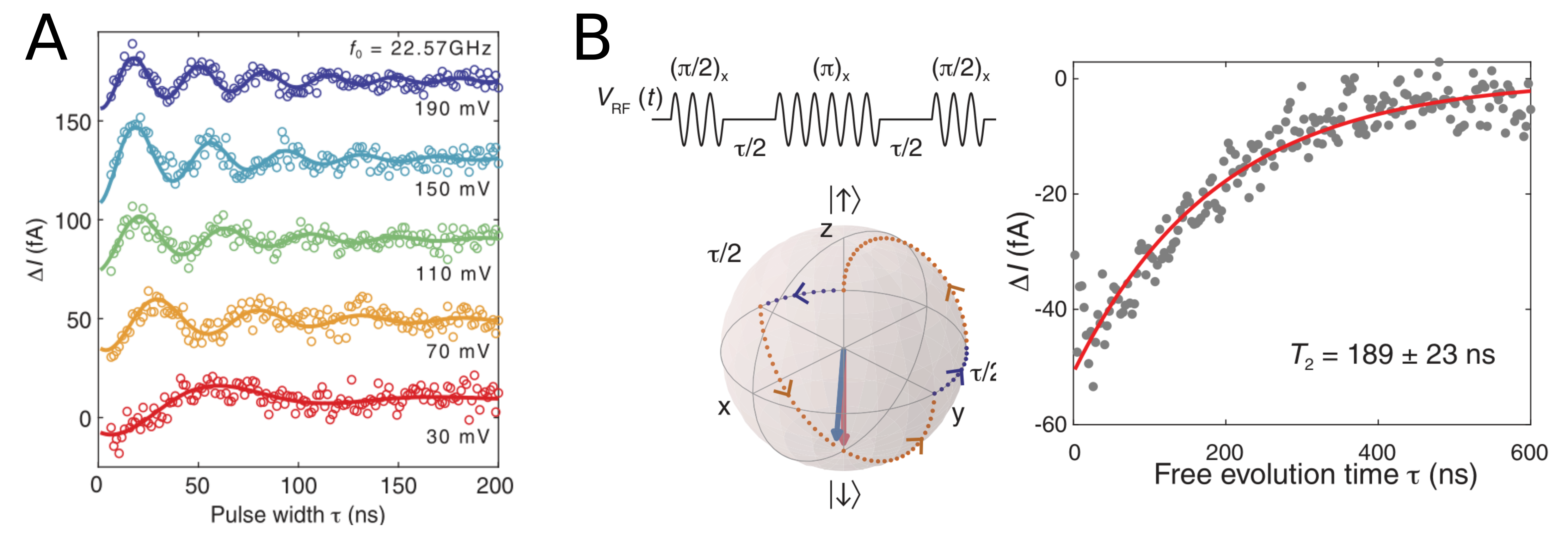
Fig. 5: A, Rabi oscillation measurements taken on a single Ti atom for different RF voltages, leading to different Rabi rates. B, Hahn Echo scheme, movement on the Bloch sphere and actual Hahn echo measurement [1].